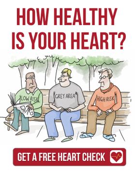
The traditional approach to heart attack risk assessment has involved the evaluation of factors that are associated with increased risk of a heart event. Among these traditional risk factors are age, smoking, diabetes, and cholesterol levels. Nevertheless, these risk factors are associations that have been identified based on large subject populations.
In other words, their predictive value pertains to an estimate of probability within large populations of people based on variables that may or may not have predictive value for the individual.
Fortunately, CT imaging is a relatively new method that is showing considerable promise in terms of predicting heart events in individual patients, which allows cardiologists to treat the risk before an adverse heart event. CT coronary angiography, a procedure that may involve the injection of contrast to gain detail about the structure of individual plaques, provides an opportunity to make an assessment that relates to plaque-specific risk, and therefore, it has important merit as another diagnostic tool in cardiology.
Regardless of a patient’s cholesterol levels, the amount of exercise he or she undertakes (or not), or how healthy his or her diet is, I want to know the health of that individual’s arteries, not the risk that a population of people with the same characteristics may have. To do this, I believe, we get the most information by using CT to look directly at the arteries.
While there is substantial data that supports traditional methods of calculating risk, holistic heart evaluation to me means that it should be combined with CT imaging together with the other traditional predictive measures, such as blood pressure and blood sugar levels. The approach needs to be about the entire patient, their situation and their needs.
In much the same way that traditional predictors of a cardiac event have some limitations, just because a patient may have low-risk based on CT imaging measures, it doesn’t mean we can ignore elevated blood sugar levels or high blood pressure, cholesterol, triglycerides and lipoproteins. The individual plaque (or plaques) may have a clear and specific impact on the risk of the potential development of a major adverse coronary event but, importantly, the risk is plaque-specific and may not necessarily match up with the risk suggested by the traditional risk factors. Consequently, it is important to comprehend that both methods provide important diagnostic clues about heart health, and they should be used in combination to determine decision-making about ongoing care and risk management of individual patients.
It is a balance between the environment of plaque formation (traditional risk factors) and local factors (what we see in the arteries).
In my opinion, the ability to combine the traditional diagnostic evaluation of an individual patient with the imaging of his or her arteries allows the most comprehensive risk evaluation for an individual, not only for the immediate future, but also for possible longer-term cardiac problems. Cardiac CT imaging will lead to a conclusion that the features observed on the scan could be low-risk features, intermediate-risk features, high-risk features or very high-risk features. This information can then be measured against traditional risk calculation variables to achieve the most reliable prediction of an individual patient’s future heart health, and most importantly, to ensure that the patient follows a treatment regimen that best reflects both their present and future heart health.
I encourage you to discuss the potential role of Cardiac CT for your health care with your local doctor.
BW - Dr. Warrick Bishop


