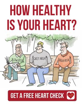
Welcome to Dr. Warrick's podcast channel. Warrick is a practicing cardiologist and author with a passion for improving care by helping patients understand their heart health through education. Warrick believes educated patients get the best health care. Discover and understand the latest approaches and technology in heart care and how this might apply to you or someone you love. Hi, my name is Dr. Warrick Bishop and I'd like to welcome you to my podcast and videocast station. And today I've got with me Dr. Karam Kostner, but we're going to do something a bit different. I'm going to introduce you first, Karam. Hi, welcome. Thank you. Look, my name is Karam Kostner. I'm a cardiologist in Brisbane and a good friend of Warrick's. So my interest is in general sort of cardiology and preventive cardiology and especially in lipids and all sorts of things, cholesterol. So today we're going to turn it around and the last couple of times Karam and I have caught up, I've taken the opportunity to interview and learn from him and share that with you. But today Karam has offered to interview me and that's because we're dealing with a pet topic. So over to today's interview, Dr. Karam Kostner from Queensland. Thank you. Thank you, Warrick. And thank you for agreeing to answer some of my questions and the questions of a lot of your listeners. And as we all know, what has really changed the game in the last 10 or 15 years in cardiology is imaging. We can now look at arteries directly and we can do all sorts of things that help us determine a patient's risk. So my first question to you is, why do we image in general? Why do we want to know about our patient's arteries? Well, there's two reasons, I think, principally, Karim. One is for you and I as a clinician, I think you'd agree it's absolutely gold to have an idea of exactly what's going on in someone's arteries when you're setting out on the journey to care for them and try and prevent problems for them in the future. The difference between someone with clear arteries versus someone who's got a lot of plaque buildup in their arteries. Your approach, your concerns, your strategies are completely different. So for you and I, in my opinion and my experience, I can't imagine a world without looking at someone's arteries to help guide our intensity of therapy and what we want to do for that person. But the thing that's really striking, Karim, is that the patients I see want to know. They really, really want to know what's going on in me, doctor. some information about the individual sitting there in front of us. No, that's excellent. And I perfectly agree with you. That was a very nice summary. What sort of imaging can we do in 2020? I mean, what sort of imaging do you use in clinical practice? Give us a couple of examples. So people will have heard of using CT for imaging the heart and we can do CT images of the heart. I'll just point out that the really exciting thing about that is, of course, that the heart moves. And because the heart moves, it's a bit of a blur to take pictures of. So it's really only been in the last decade where broadly we've had technology advanced enough to allow us to freeze the heart and take really good pictures that reliably and reproducibly give us a good idea of what's going on in their heart. Well, when we use a CT, we can take pictures without contrast injection. And then we're really looking principally for calcium as a marker of plaque in the arteries, but we can also inject contrast. That gives us not only the calcium, but can show us plaque, plaque composition, whether there's narrowings, how big are the arteries that are involved, and even more detail above that to help us guide our therapy. The other sort of technology we can use to image people, as you're aware, is ultrasound. And ultrasound is a non-invasive, non... radiation based technology where we can image arteries that are close to the skin like the neck or the femoral arteries looking directly for evidence of plaque but you know as well as i do we can use ultrasound even for clever things like looking at the achilles tendons for buildup of cholesterol in a particular patient set where people have really high cholesterol levels so it's a little bit of imaging we can do to keep us interested Exactly. Now, most of our listeners are familiar with an echocardiogram with an ultrasound of the heart that looks at the heart muscle and valves. And a lot of our colleagues talk about functional versus anatomical imaging. And that's quite complicated sometimes for our patients. Can you explain a little bit what the difference is and where you would do functional imaging versus anatomical imaging? Or do we need both? Yeah, no, that's a great question. Look, functional, I often try and discuss or use car analogies when I speak with my patients. And I think a car analogy allows us to all relate because most of us have driven cars around or understand in the simplest way, the way a car engine works. Well, very simply a functional test for your car to test its fuel lines. And really we're talking about fuel lines of the heart, the coronary arteries, a functional test of the fuel lines. for your car would be to drive it up one of the steepest streets in your neighborhood and see if the car coughs or splutters or shows features of lack of fuel getting to the engine. Well, we do that with a stress test. We put people through a stress. We run them literally up a hill and see if the heart shows features of lack of blood flow. Well, the flip side of that is exactly what you said, which is an anatomical test. Imagine instead of testing the fuel line of your car, you could Literally take an X-ray of it and see if there's any stuff clogging up or building up within that fuel line if it's going to cause a problem in the future. And it's exactly the same with our hearts. We can use a CT scan to take pictures of the arteries and look for rust in the pipes before it's even causing a problem. Very good. Very good analogy. And that's one that's easy to understand. Now, the next important question when we talk about imaging is. Who do you image? Do you image everybody, for example, with high cholesterol, with a CT or calcium score? Do you do a stress echo on young people who have symptoms for cardiovascular disease? Who do you image in your practice? So, look, I think this is really important. And I have to say that I see this, it appears to me, being poorly applied broadly within our own specialist group, Karam. And I see... patients being sent for functional testing. And these are patients who may have no symptom whatsoever. They're perfectly well. Like you and I, we have no symptom. We can undertake activities without any issues at all. To put you and I through a functional test to see if our pipes are narrowed makes no sense. So you and I would be perfect candidates for imaging to assess our arteries. because the chance of picking up something abnormal on a functional test is incredibly low. So in general terms, I use functional testing or stress testing when someone presents with a symptom, which is different to a risk assessment. In risk assessment, I think there are a group of patients who really can benefit from imaging. For those people where you're looking at risk assessment, Generally, they have to be of a certain age. You don't start doing this on teenagers. That just isn't standard. But for the average cohort, I generally think men around 50 years of age and women around 60 years of age are really good candidates for having a conversation about imaging. Is that sort of where you see it yourself? Yeah, I agree, especially if they have risk factors, you know, if they sort of have a family history or high cholesterol or high blood pressure or a combination of those, yeah. So one of the things that I try and do is I incorporate the Australian cardiovascular disease risk calculator or similar risk calculator to get an interpretation of what population group that this individual would sit in. So imagine I take a 50 year old male with average blood pressure, average cholesterol, and we see that they've got a risk of event over the next 10 years of say 10%. recommendations for treatment, they're a little bit ambivalent, and there's an opportunity for the doctor and the patient to decide whether intervention or not, or supervision or lifestyle modification is appropriate. Well, I think in that sort of situation, imaging provides a fantastic opportunity to be more precise, because if we think about that risk of one in 10, what it's actually saying is if we take a population of 100 men with the same characteristics and follow those 100 men for 10 years, and then we'll have a heart attack. Well, imaging allows us to look at the whole population and find the 10 or 20 that look really high risk where those events are likely to come from. And that's when we can target therapy and be precise about it. Now, that makes sense. And since you're talking about imaging with CT, for example, what is the difference between a calcium score and a CT angiogram? So I touched on that a little bit earlier. When we do a calcium score, we don't inject any contrast at all. We're simply taking images of the heart and looking for calcium as the marker of plaque, nothing else. Generally, we can simply add, well, in my own practice, I tend to add a CT coronary angiogram to calcium score if there's calcium present to get more detail around the plaque. And that's an injection of contrast that we literally track through the body, it passes through the arteries of the heart during that time we take a picture but it outlines not only the inside of the arteries but the wall of the arteries as well so it shows us plaque in development not just light plaque Okay. And from what age can you do a coronary calcium score, for example, in your practice? I mean, young people with vascular disease, surely 20, 15, 25-year-olds wouldn't qualify for a coronary calcium score. We often do carotid ultrasound on these young individuals. You know, we look at the intima media thickness on the neck arteries, etc. But at what age do you usually start with cardiac CT? how severe their cholesterol levels are. So if they're moderate or intermediately elevated, then we can have a conversation about imaging. If they're very, very elevated, as in severe familial hypercholesterolemia, then I would make a strong advocacy that all our research would support treating those people regardless is a good thing in the longer term. So probably the youngest people I would... Look to image would be males at 30 odd years of age. I wouldn't generally go any lower than that. Women at 40, but these are only in the situation of very severe family history, possibly other risk enhancers like lipoprotein little A elevation, possibly some hypertension, possibly pre-diabetes, a very adverse lipid profile, things that would really raise concern. Yeah, that's what we do as well. And do you do serial imaging in those people? Or do you do it once and then sort of treat? Or when do you do the test again? Let's put it that way, especially if it's normal. So if it's normal, there are some guidelines. The Americans in their prevention guidelines from 2019 suggested that we can repeat this sort of testing in the average population who have a zero score at about five years. And I think that's probably not a bad... guide. You can imagine though that that isn't across the board because it varies depending on the situation or the context. Imagine if we scanned a gentleman who is 85 years of age, he had a zero calcium score, then there's no way that he would have a meaningful repeat test of five years. He's already defined his natural history to a large degree. So age, sex and other risk factors come into it, but five years is a good roundabout figure, I think. Very interesting. The next question I have, Warrick, is what's involved in this test? Is it complicated? Do people have to do something special? Or how does it look in practice? In my own practice, Karam, I'm very strong about seeing the patient, explaining the test to them. Currently in Australia, cardiac CT imaging, either calcium scoring or CT coronary angiography is not. a guideline recommendation in Australia. Although we've got a position statement on it written by Cardiac Society and our friend Christian Hamilton Craig, or led by Christian Hamilton Craig, and although the Heart Foundation of Australia are writing a position paper on it, we don't have a formal guideline recommending it. In that situation, I'm really clear with patients that they need to be informed and know that it's a test that they want to proceed to. I'm happy to support it as long as they understand. There's no Medicare rebate. I like to explain that to patients. I really like to explain to patients the radiation dose, which is not very high. And I know you're probably going to ask about it, but I'll jump in early. The radiation dose for a calcium score and for a CT scan run very close to what we call in radiation terms, one millisievert. But in practical street terms, that's about the same as a mammogram. So you can get two scans of your heart. for about a mammogram for each stage of that scan. So two scans, two mammograms. And lastly, because my patients will often proceed to injection of contrast if they have calcium present, I like to explain that there is a small but real risk of an allergic reaction to that injection of contrast. It's about one in 200,000. So it's not a lot. And in fact, Just for a bit of trivia, it's three times safer than peanut butter, which has a one in 70,000 catastrophic allergic response. And we find that on supermarket shelves. But nonetheless, it's really important to let people know that. I also let people know I don't inject contrast if there's a zero calcium score. I don't think the evidence supports that. So personally, I want to speak with the patient, inform them, make sure they fully understand what's going on. And then I give them heart rate regulating medications. They turn up on the day, often fasted with beautiful heart rate because I've given them tablets to control their heartbeat. The test is very straightforward. It's a breath hold and acquisition of images. And if a contrast is required, someone will pop a little drip in the arm, which is really a fairly non-invasive procedure. And they may get a hot flush feeling from the injection of contrast. And that's it. Very useful information. When you say the coronary calcium score is not reimbursed, it's the same in Queensland. You know, patients pay between $120 and $150 for it. But I have to say, if the question is chest pain and we do a CT angiogram, it is Medicare built, at least in Queensland. It'll probably be the same in your setting. Yeah, well, that's an evaluation of someone with symptoms. A little bit different to where I was talking about. risk assessment. But yeah, no, completely right. Yeah, I understand. And then the other question I had is, can anybody do a CT angiogram or a CT coronary calcium score and report it? Or is it usually cardiologists or radiologists? Just for our listeners, obviously. Well, Karam, I know you're probably poking the bear because you know this is a bit of a soapbox for me. I've personally reviewed... somewhere close to 3 000 patients where i've seen the patient sent them for the scan after explanation i've personally looked at their images and i've personally interpreted the risk i believe inherent and then match that up with the individual patient's characteristics to find what i believe is the best solution for them in conjunction with their needs wants wishes and tolerability of different medications so i think it's a highly specialized area I agree with you. And that's why I'm asking. I think it's important to get that message across. You know, I think very similar to you. As you know, I get very distressed when I see this fantastic tool being poorly used by people who don't fully understand it. And I have no issue with people asking for help or assistance, but I do get disappointed if... A colleague does one or two here or there, really doesn't understand what they're doing. And potentially the patient may be going through a test, paying for the test and not having it interpreted in the way that is best for that individual's outcome, which says to me, why would you bother in the first place? Understand. Now, in the remaining couple of minutes, and that was very useful, I've learned a lot as well, Warrick, I would like to talk about two things. And one of them is when not to do this test. And especially CT with contrast, for example, is the situations where patients should not have that test. So, for example, people with impaired renal function, people who are in very fast atrial fibrillation where the heart rate is not low, et cetera, et cetera. Yeah. Look, that's... That's a very straightforward and important question. We don't do it on everyone. There's no test that's perfect for everyone. Like there's no medication that doesn't have a side effect. Like there's no magic bullet for almost anything. So if you've had a heart attack or a stroke or bypass grafting or stenting, then doing CT imaging for risk is altogether wrong. Occasionally we would do CT imaging. but very specific situations if you've had a problem with your heart before. So that's probably the most important mistake that people actually make. And I've seen that done. You're exactly right. You wouldn't give someone with renal impairment contrast and heart rate control is really important. But other than that, there's not really too much in terms of contraindications. Of course, if there's a contrast allergy, you would avoid contrast. So that contrast history is really important. And age, I think is important for someone who's really young. I really try and avoid using CT imaging because of the radiation. I tend to err towards ultrasound of the carotid arteries. Very good. And the last question I had, is there some alternative methods that are being developed at the moment that we should at least, our listeners should have at least heard about? such as MRI of arteries, such as PET scan, et cetera? Or is it really all about CT and echo and ultrasound at the moment? Yeah, for looking at the arteries in the way that we're talking about, which is a predictive risk stratification for future likelihood of event based on plaque burden, CT is way out in front. And that's really because of the temporal resolution, which means CT can acquire these images really quickly. MRI isn't quite fast enough to capture the arteries. And so we just don't get enough of the detail down the length of the artery. We can get good origins of the arteries in MRI, but CTs really got it for non-invasive rapid acquisition. There is some stuff with PET scanning, which is in research. facilities at the moment where they're looking at uptake of markers to show inflammation within the plaque and that may be something that's a decade or two away but it's remarkably interesting to think that we might be able to show spots within the arteries that could actually be the problems where a rupture occurs because there's inflammation and activity occurring but we're a long way from that in a practical sense right at the moment. Excellent. Well, that's all the questions I had, Warrick. That's fantastic. We've done about 20, 25 minutes. I'll hand back to you to close this podcast. But that was very informative, even for me who deals with these things every day. Karim, thank you so much for interviewing me today. For those listening, I hope you got something out of it. If you have any queries or questions, please drop us a line at info at drWarrickbishop.online if you've got any suggestions. For future podcasts, of course, let us know. Until next time, please keep well and please don't die from a heart attack. Goodbye. You have been listening to another podcast from Dr. Warrick. Visit his website at drWarrickbishop.com for the latest news on heart disease. If you love this podcast, feel free to leave us a review.